TMA CONTROL SOFTWARE
he TMA Control software is the perfect solution for TMA block design and creation.
Up to a couple hundreds of samples can be fitted on a single slide and all these samples are stained at the same time. The evaluation and scoring of several hundred samples is impossibe by tradtional microscopy.
Tissue microarrays represent a significant move towards economy and constant quality in immuno-histochemistry: up to a couple hundreds of samples can be fitted on a single slide and all these samples are stained at the same time. Little arrays are taken out of the specimen with a needle-like tool and then aligned in blocks. After sectioning these blocks are put on a slide and stained homogeneously.
TMA is useful when the doctor is only looking for signs of specific parts (proteins usually) in the sample. With the means of immunohistochemistry it is possible to detect and to visibly differentiate the desired proteins which means it can be effectively used e.g. in cancer diagnosis. It is a very rational step to fit as many samples (here: cores) on one slide as possible because staining in constant quality is essential in immunohistochemistry. However, this creates a problem traditional microscopy cannot cope with efficiently.
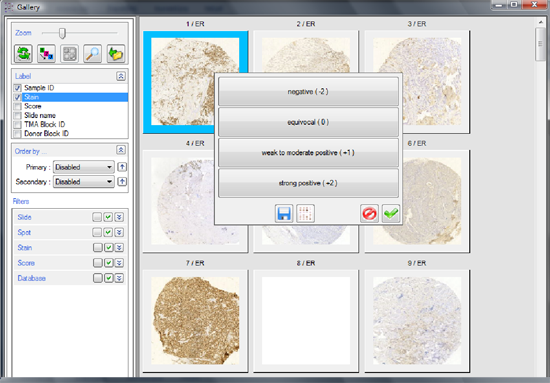
The much greater amount of samples requires much greater administrative work. You have to find the core, record your score and then move to the next position. It is easy to miss. If you want to compare your previous scores with your present ones it is almost impossible because navigation in the traditional microscope was not invented for this kind of job.
This is one of the fields where virtual microscopy clearly shows its strengths. 3DHISTECH Ltd is very proud to present its TMA software module to you. With the TMA module of Pannoramic Viewer you can enjoy all the benefits of tissue microarrays.
he TMA Control software is the perfect solution for TMA block design and creation.
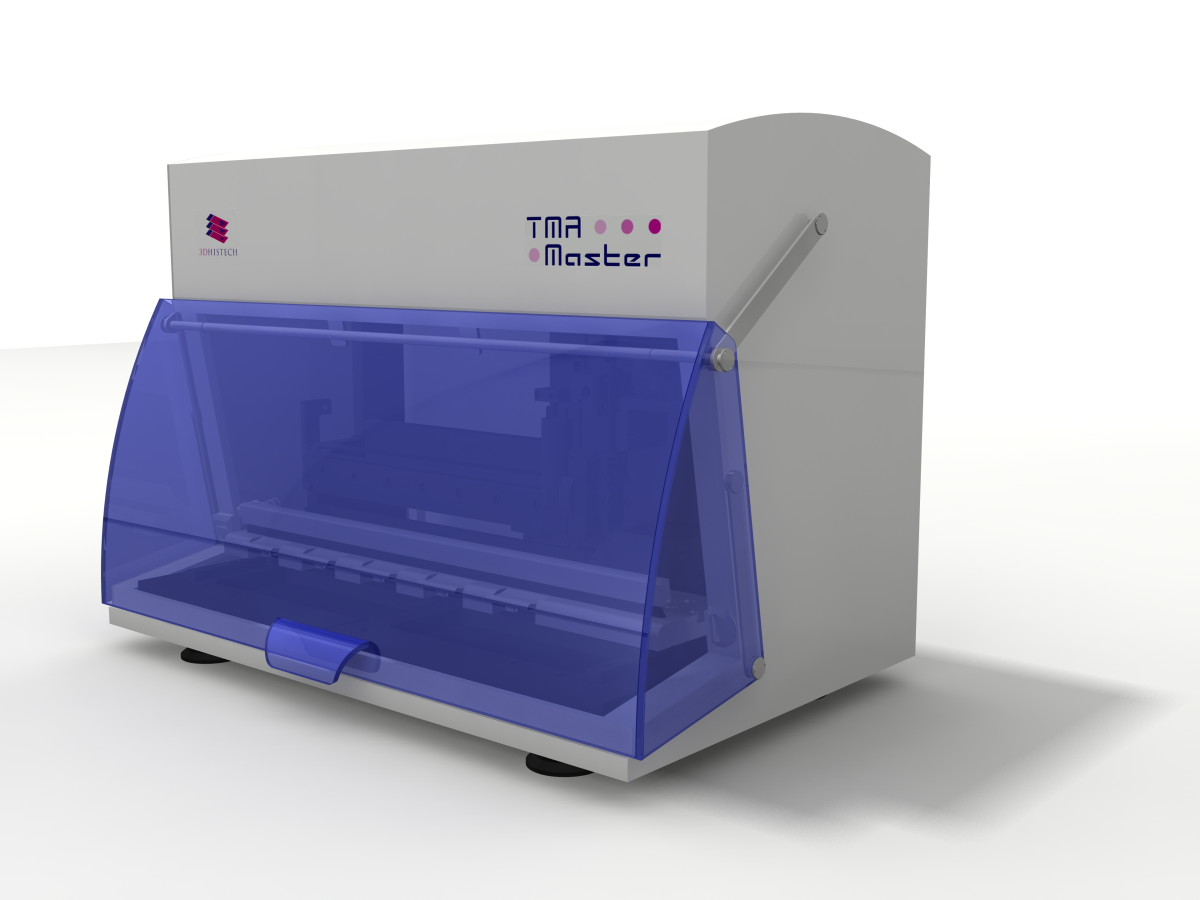
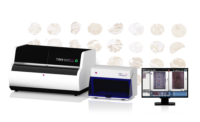
TMA Master II is the smallest fully automated tissue microarrayer at the market, it easily fits on any laboratory bench.

The TMA Grand Master is the fastest, highest capacity and most user friendly tissue microarrayer on the market.