CASE CENTER

CaseCenter

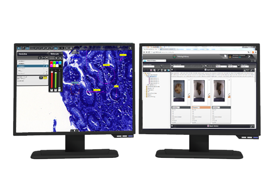
CaseCenter is a powerful, server-based slide management system with a fully featured slide database capable of storing both macroscopic images and digital slides. Thanks to its flexible structure, it can be adapted to various fields, including clinical pathology, research applications, teleconsultation and education.

Consultation plays an ever growing role in pathology. Faster and better diagnosis requires faster and better access to the medical samples. Digital slides make this all possible. CaseCenter, as a full featured whole slide image management software with teleconsultation abilities, helps in the process!

CaseCenter can be used in smaller or bigger laboratories or even hospitals or hospital networks. It can be integrated into existing hospital information systems.
CaseCenter is a flexible, distributed server system: more servers can easily be added for enhanced data security and storage requirements.

Licensing is done on the server side: CaseCenter regulates the number of users who can access the server at any time. User & group rights management is built in the system.

Structure

HUB: web front end, MySQL database Service Unit (SU): slide storage

Automatic slide alignment

Automatic and rapid alignment of series of sections even with different stainings. It is not necessary to rotate digital slides manually.

Virtual Slide and VirtualTray

All slides of a case represented in one view by one single click for faster case reading and slide comparison. Combine multiple digital slides or regions of interest into one VirtualTray without the need for more storage!

TMA workflow support

from target marking up to the core visualization. Use CaseCenter’s remote slide access for sample designation or TMA slide evaluation: fast, convenient, and easy.

Transparent login method for high traffic user authentication

Login queue and automatic logout. It is possible for the administrator to log users out after a specified time of inactivity. A login queue handles the shortage of free licenses. Users get in the queue according to their user levels (users with higher rank are put forward in the queue) and their queue number (position in the queue) is displayed and is automatically updated.

Server side barcode parsing

Automatically create folders and Cases from the barcode on the slide on server side Advantages:

  • No need to define path to every slide. Slides are put in their place automatically by barcode
  • It also works with manually written indentifiers
  • Recognizes and uses cases/folders created by HIS
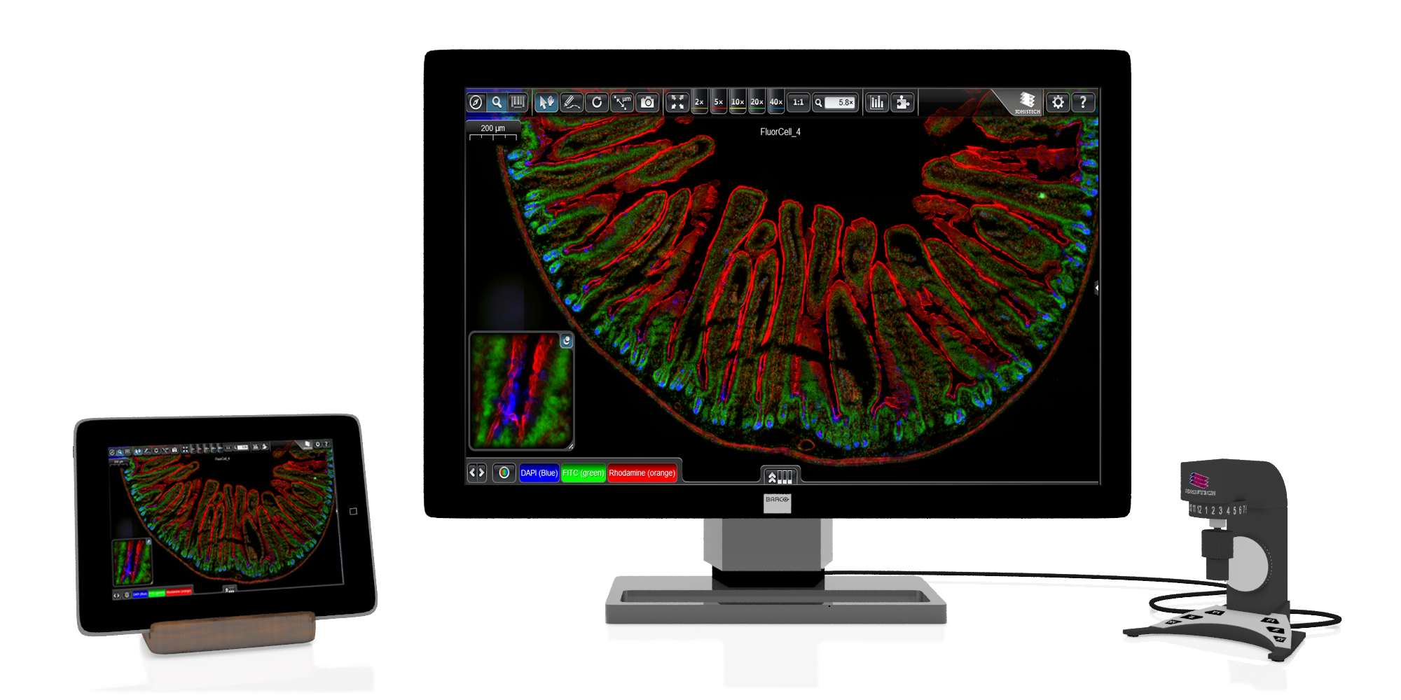
InstantViewer

New, multiplatform slide viewer application Supported platforms: Windows, Win Mobile, Mac OSX, iOS, LINUX, Blackberry

Secondary Service Unit

Using a secondary Service Unit when the default Service Unit is full or unavailable or getting errors

  • Quick search in folder tree
  • The uploader of each slide is shown in slide details
  • "Delete folder” and "Delete slide” functions are set to user level (available only for a pre-defined user level and above)
  • Send daily e-mail about the status of the Service Unit: Send a message to the admin every day about the condition of the Service Units
  • If a Service Unit becomes unavailable and it is not available for a certain time, a message is sent to the admin. This e-mail contains the name and current status of the Service Unit.

New features

  • Redesigned Graphic User Interface: This new GUI represents the new common GUI concept of all 3DHISTECH software applications such as the CaseViewer 2.0 in order to harmonize the different applications, create synergies and make them “easy to use”
  • Customizable application header supports the possibility of adding a logo of the user institute.
  • Reporting during teleconsultation: During a teleconsultation session a mutual case report can be created by the participating parties or an already created Case Report attached to the Case can be shared.
  • Case Reports can be created and attached to a Case including one or more digital slides
  • Case Sharing: similarly to existing “Slide Sharing” function, a case (a set of digital slides and attached reports) can be shared with other users or with groups of users
  • In case of eg. “second opinion” a consultant can add his personal report to a shared Case by using the HTML based Report Templates (Slide Manager / Teleconsultation / Share). The saved report will be converted to PDF.
  • Improved algorithm for slide alignment

More features

  • Web based slide and case database with fast search
  • Teleconsultation support with Pannoramic Viewer
  • Easy extension by adding new SUs
  • Slide access through the free Pannoramic Viewer, the InstantViewer or the iPad Viewer
  • Virtual Slide handling
  • MS Network, HTTP and HTTPS accessibility
  • Fully manageable user and user group rights system
  • Direct scanning into database with Pannoramic digital slide scanners
  • Macro images and multiple file attachments stored in the case
  • CaseCenter can work with other 3DHISTECH software applications (e.g. quantification software can be run on remote slides)

Minimum requirements

  • Processor: Intel® Xeon® Processor E3-1220
  • 4 Core CPU
  • 16 GB RAM
  • 320 GB + slide storage: depends on requirements
  • Slides: 0,5-1 GB / slide
  • MS Windows Server 2012 R2
Recommended Network Bandwidth
Scan1, Scan2, … Scan n 1 Gb/s
View1, View2, …. View n 10/100 Mb/s
SU1, SU2, … SU n, SAN (in case of 1 scan) At least 1 Gb/s
SU1, SU2, … SU n, SAN (in case of multiple scan) 10 Gb/s
Wan3 (remote viewer station) At least 500 kb/s for every concurrent viewer sessionb Optimal 2-5 Mb/s for every concurrent viewer session
Wan1, Wan2 (Broadband access) At least 500 kb/s for every concurrent viewer session Optimal 10-20 Mb/s
Storage capacity planning (Service Unit / Storage)
Avg tissue single layer with 20x (15mmx15mm)~ 1GB
Avg tissue single layer with 40x (15mmx15mm)~ 2,5 - 3GB
Avg tissue with n x Z-layers n x single layer GB
CaseCenter site requirement, CaseCenter Server (SU / HUB)

Tissue Classification

Colour- and pattern-based image analysis solutions are used to identify different tissue elements in stained tissue. Instead of the time-consuming manual annotation, automated detection and annotation of a region of interest is available with a quick training on relevant tissue samples.

HistoQuant

HistoQuant is a histological segmentation module which identifies relevant histological structures or tissue elements based on the colour and intensity of the image pixels. This module can analyze fluorescent and brightfield slides. Furthermore, the co-localized pixels can also be detected in double-stained tissue.

PatternQuant

PatternQuant is a trainable pattern recognition module for tissue classification, tissue pre-segmentation and identification of several tissue structures. The artificial intelligence-based algorithm is able to learn and classify various tissue types based on their texture patterns and colour features.

IHC Quantification

A special IHC colour calibration option is integrated into these quantification modules, facilitating software integration to the local laboratory. By applying this function the software can be calibrated to the sample quality (local laboratory stain protocol, or different stainer), resulting in an adequate measurement.

NuclearQuant

NuclearQuant is designed for cell nuclei detection and quantification of IHC stained samples. NuclearQuant measures several morphological features besides stain intensity. The cell nuclei classification and the final score are calculated by the intensity score and the proportion score.

This module has IVD approval for the analysis of Estrogen- and Progesterone-stained breast tissue samples.

MembraneQuant

MembraneQuant is a cell membrane detection software application that can be used for the quantification of IHC stained histological samples. This module measures cell morphology and stain density, reports intensity-based core ranges, overall scores and positivity percentages (including H-Score), distinguishing the continuous membrane stain from the moderate stain.

This module has IVD approval for the quantification of Her2 expression in breast tissue samples.

CellQuant

CellQuant is a cell detection application, an optimal solution for investigating various IHC stainings. The application is adequate for cell nuclei, cytoplasmic and membrane marker quantification. The software reports the results like positivity ranges of cell nuclei, cytoplasm or membrane signals.

DensitoQuant

DensitoQuant is an easy-to-use, fast and accurate, stain-intensity-based IHC quantification tool. The application identifies the positive stain, based on an automatic colour separation method through which individual positive pixels are counted and classified based on intensity and threshold ranges. So finally an H-Score can be calculated based on the proportion of the positive and negative pixels.

Molecular Pathology

Fluorescent in situ hybridization (FISH) and Chromogenic in situ hybridization (CISH) are routinely used methods in histopathological diagnostics. The visual evaluation of these samples is a difficult task, and typically a time-consuming process and prone to human errors that can be eliminated by using image analysis solutions.

FISHQuant

FISHQuant is a powerful cancer and cytogenetic application dedicated to quantifying FISH (Fluorescent In Situ Hybridization) signals on tissue samples of solid tumour diseases like breast cancer, lung cancer, sarcoma symptoms, and lymphomas. This module is suitable for examining haematological tumours. FISHQuant classifies the interphase and metaphase cells individually for an even more comprehensive evaluation. This module has IVD approval.

CISHQuant

CISHQuant quantifies CISH (Chromogenic In Situ Hybridization) stained samples. The algorithm can be calibrated to the staining protocol and quality by using an integrated colour setting tool. This module is a suitable module for examining gene amplification, deletion and chromosome aberration.

Batch Analysis

Using the batch analysis mode, multiple digital slides can be examined in the background, saving a significant amount of time. Measurements can be run after slides, annotations and profiles have been selected. All measurement results can be exported into an Excel file.

Data Visualization (DVT)

An integrated data visualization mode is available for each module. The result can be visualized on a table, scatterplot, pie chart or a histogram by using this feature. There are integrated galleries where the detected and classified cells can be presented.

Product brochure

Tutorials

Her2stain quantification using PatternQuant and MembraneQuant

Breast Cancer Tissue Analysis using PatternQuant and NuclearQuant

RELATED PRODUCTS

CASEVIEWER

CaseViewer is designed for supporting histopathological diagnostic workflow

INSTANTVIEWER

Open and share your slides with the multi-platform InstantViewer!